Anatomical Name Of Lower Back Muscles : Muscles Of The Lumbar Spine Of The Trunk : The extensors, which include the many muscles that attach to the spine and work together to hold your back straight while enabling you to extend it.
Anatomical Name Of Lower Back Muscles : Muscles Of The Lumbar Spine Of The Trunk : The extensors, which include the many muscles that attach to the spine and work together to hold your back straight while enabling you to extend it.. The lats are attached to the upper end of the humerus with fibers running down in a fan down the vertebral column and pelvic girdle. Anatomy of back muscles 12 photos of the anatomy of back muscles anatomy of back muscles in. There are three parts to the trapezius. The spine's four sections, from top to bottom, are the cervical (neck), thoracic (abdomen,) lumbar (lower back), and sacral (toward tailbone). Vastus medialis, vastus lateralis, vastus intermedius, rectus femoris comments:
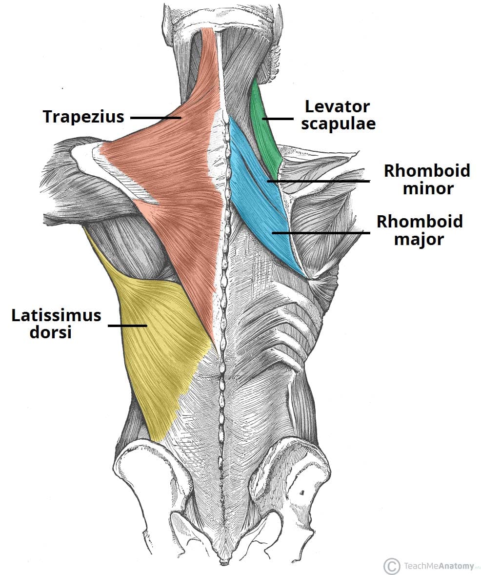
The anatomy of the back muscles the back is subdivided into the upper, middle, and lower back. It is composed of trapezius, latissimus dorsi, rhomboid major, rhomboid minor and levator scapulae. The abdominal and lower back muscles work together to form a supportive girdle around your waist and lower back. There are three parts to the trapezius. Related posts of muscles of the lower back and buttocks diagram muscle anatomy of the thigh.

The muscles of the lower back, including the erector spinae and quadratus lumborum muscles, contract to extend and laterally bend the vertebral column.
Related posts of muscles of the lower back and buttocks diagram muscle anatomy of the thigh. By the way, have you heard about the myth of. Anatomy of back muscles 12 photos of the anatomy of back muscles anatomy of back muscles in. It starts all the way down in your lower back, climbs up to the middle of your back, and stretches out into your shoulder. Intermediate extrinsic muscles of the back: Lumbar spine anatomy video save the lumbar region of the spine, more commonly known as the lower back, is situated between the thoracic, or chest, region of the spine, and the sacrum. Opposite this, on the back of the leg, is the hamstring muscle. Located at the front of your body, the flexors. Hamstrings (semi membranosus, semiteninosus, biceps femoris), gastrocnemius, popliteus extensors: The spine's four sections, from top to bottom, are the cervical (neck), thoracic (abdomen,) lumbar (lower back), and sacral (toward tailbone). The back muscles represented on an anatomical chart and on a schematic view of the origin and insertion of the proper muscles of the back (iliocostal muscle of. Sherwin is a medical research scientist and author of the low back pain program and ebook. This article looks at the anatomy of the back, including bones, muscles, and nerves.
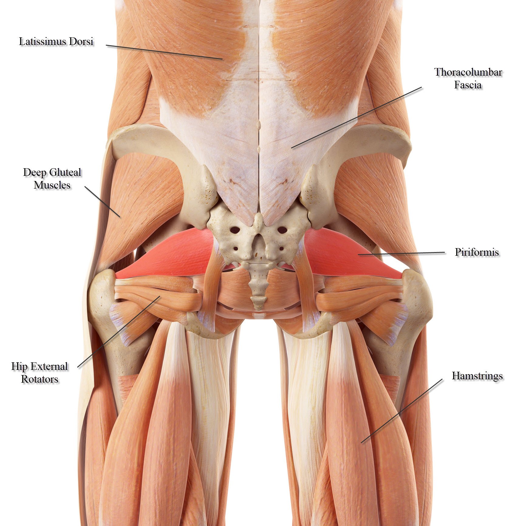
Working our way down the lower body, the first muscle group is the glutes (including the gluteus maximus, medis and minimus), otherwise known as the buttocks. Three main muscle groups are located in the lower back: The main movements of the knee are flexion and extension.for lateral knee pain, look at the vastus lateralis. This bone is shaped like a triangle that fits between the two halves of the pelvis, connecting the spine to the lower half of the body. This article looks at the anatomy of the back, including bones, muscles, and nerves.
Working our way down the lower body, the first muscle group is the glutes (including the gluteus maximus, medis and minimus), otherwise known as the buttocks.
The pelvic floor muscles also help increase this pressure, which provides stability to the spine and trunk. The muscles of the lower back, including the erector spinae and quadratus lumborum muscles, contract to extend and laterally bend the vertebral column. Leaning back to straight vertical and all points in between. Muscle anatomy of the thigh 12 photos of the muscle anatomy of the thigh anatomy of the anterior thigh muscles, muscle anatomy of upper thigh, muscle anatomy thigh mri, muscles of the leg grey's anatomy, muscles of the thigh ct anatomy, human muscles, anatomy of the anterior thigh muscles, muscle. The lats are attached to the upper end of the humerus with fibers running down in a fan down the vertebral column and pelvic girdle. The trapezius or trapezoid muscles are two paired muscles that extend from the base of the thoracic vertebrae in the spine to the occipital bone and run out to the spine of the scapula. Working our way down the lower body, the first muscle group is the glutes (including the gluteus maximus, medis and minimus), otherwise known as the buttocks. These muscles provide posture and stability to the body by holding the vertebral column erect and adjusting the position of the body to maintain balance. Attached to the spine by soft tissues call tendons, these muscles control back motions, support the spine and enable you to stand, bend, twist, walk and and move in different directions. Muscle structure of the lower back. Related posts of muscles of the lower back and buttocks diagram muscle anatomy of the thigh. The muscles of the back with the surface (trapezius, latissimus dorsi, thoracolumbar fascia, deltoid) and intermediate layers (serrated muscles, external and internal oblique muscle). The sacral region (bottom of the spine) below the lumbar spine is a bone called the sacrum, which makes up the back part of the pelvis.
These muscles provide posture and stability to the body by holding the vertebral column erect and adjusting the position of the body to maintain balance. Extrinsic and intrinsic.the back functions are many, such as to house and protect the spinal cord, hold the body and head upright, and adjust the movements of the upper and lower limbs. The flexors, which attach at your lumbar spine (lower back), and enable you to bend forward. The two main muscle groups involved in back function are: Related posts of muscles of the lower back and buttocks diagram muscle anatomy of the thigh.
The two main muscle groups involved in back function are:
Sherwin is a medical research scientist and author of the low back pain program and ebook. The superficial group, also known as the appendicular group, is primarily associated with movement of the appendicular skeleton. Shoulder muscle anatomy neck muscle anatomy anatomy back neck and shoulder muscles gross anatomy lower back muscles anatomy female back muscles female torso chest muscles. Leaning back to straight vertical and all points in between. The sacral region (bottom of the spine) below the lumbar spine is a bone called the sacrum, which makes up the back part of the pelvis. The back muscles represented on an anatomical chart and on a schematic view of the origin and insertion of the proper muscles of the back (iliocostal muscle of. The lats are attached to the upper end of the humerus with fibers running down in a fan down the vertebral column and pelvic girdle. Lower back muscle anatomy includes the multifidus, longissimus, spinalis, and quadratus lumborum. All the extrinsic back muscles are innervated by the ventral (anterior) rami of the cervical spinal nerves , except for the trapezius muscle which receives its supply from the accessory nerve (cn xi) . The muscles of the lower back, including the erector spinae and quadratus lumborum muscles, contract to extend and laterally bend the vertebral column. The back is the body region between the neck and the gluteal regions. Related posts of muscle names of lower back anatomy of back muscles. Extrinsic and intrinsic.the back functions are many, such as to house and protect the spinal cord, hold the body and head upright, and adjust the movements of the upper and lower limbs.


Komentar
Posting Komentar